Loculated Pleural Effusion Cxr / Fig 1 10 Empyema As A Loculated Pleural Diseases Of The Chest Breast Heart And Vessels 2019 2022 Ncbi Bookshelf - Pleural effusion is an abnormal accumulation of fluid in the pleural space.
byAdmin-
0
Loculated Pleural Effusion Cxr / Fig 1 10 Empyema As A Loculated Pleural Diseases Of The Chest Breast Heart And Vessels 2019 2022 Ncbi Bookshelf - Pleural effusion is an abnormal accumulation of fluid in the pleural space.. In chf effusions are bilateral and more on right. Loss of right diaphragmatic and cardiac silhouettes. A right thoracentesis was performed, and on seeing the biochemistry results, the left side was also punctured. Malignant pleural effusion, breast carcinoma, maliganancy: Loculated effusions are collections of fluid trapped by pleural adhesions or within pulmonary fissures.
A right thoracentesis was performed, and on seeing the biochemistry results, the left side was also punctured. Loss of right diaphragmatic and cardiac silhouettes. Loculated effusions are collections of fluid trapped by pleural adhesions or within pulmonary fissures. Bilateral pleural effusion (bpe) is not an uncommon finding in clinical practice. Sometimes in the setting of pleuritis, loculation of fluid may occur within the fissures or between the pleural layers (visceral and parietal).
Pleural Effusion Knowledge Amboss from media-us.amboss.com Bilateral pleural effusion (bpe) is not an uncommon finding in clinical practice. Pfa = pleural fluid analysis. Loss of right diaphragmatic and cardiac silhouettes. Treatment may fail if the catheter is not placed optimally within the loculation or if the fluid is hemorrhagic or fibrinous. Weight loss 15 lbs in one month • pf is a transudate; The right pe was larger and loculated (by ultrasound). Normally, a small amount of fluid is present in the pleura. Line not corresponding to fissure
This type of effusion is empyema unless proven otherwise.
Most pleural effusions, whether free flowing or loculated, are hypoechoic with a sharp echogenic line that delineates the visceral pleura and lung. This type of effusion is empyema unless proven otherwise. We studied the value of transca … Complex septated, complex nonseptated, or homogeneously echogenic effusions are always exudates (fig. Loss of right diaphragmatic and cardiac silhouettes. The fluid buildup may be the result of a chronic condition like congestive heart failure. Pfa = pleural fluid analysis. An effusion is exudative if it meets any of the following three criteria: Case 1 • 77 year old woman with hx of copd • 2 week history of uri symptoms • zpak and then 10 days antibiotics • hospitalized with 3 day history of fever to 39.0 °c, shaking chills, nausea and large pleural effusion. 1 article features images from this case 20 public playlist include this case Loculated right pleural effusion with foci of atelectasis and consolidative changes concerning for pneumonia. A pleural effusion is a collection of fluid in the space between your chest wall and lungs. Draw out the diagram of steps and findings of pleural effusion starting with finding a plural effusion (on upright cxr or pe) when getting a theracocentesis you send out 4 tubes
The right pe was larger and loculated (by ultrasound). 1 article features images from this case 20 public playlist include this case Icu patients cannot sit up and the effusion layers posteriorly. This type of effusion is empyema unless proven otherwise. Left pleural effusion is a development of excessive fluid in the left side of the pleural cavity, the space surrounding the lungs.
Pleural Effusion Springerlink from media.springernature.com A pleural effusion is a collection of fluid in the pleural space. A right thoracentesis was performed, and on seeing the biochemistry results, the left side was also punctured. A loculated pleural effusion can mimic a mass hence is sometimes known as a pleural pseudotumour. Localized interlobar effusions in congestive heart failure (phantom or vanishing lung tumor/s) is/are uncommon but well known entities. The pleura are thin membranes that line the lungs and the inside of the chest cavity and act to lubricate and facilitate breathing. Enlarged mediastinal lymph nodes, possibly reactive. What are the different appearances of pleural effusion? Case 1 • 77 year old woman with hx of copd • 2 week history of uri symptoms • zpak and then 10 days antibiotics • hospitalized with 3 day history of fever to 39.0 °c, shaking chills, nausea and large pleural effusion.
Transudative effusions are a result of pressure filtration without capillary injury (i.e hydrostatic and oncotic pressure abnormalities).
Complex septated, complex nonseptated, or homogeneously echogenic effusions are always exudates (fig. Most pleural effusions, whether free flowing or loculated, are hypoechoic with a sharp echogenic line that delineates the visceral pleura and lung. The first step in evaluating pleural effusions is determining whether it is transudative or exudative. A chest tube (12f) was inserted under imaging guidance into the largest locule. The fluid buildup may be the result of a chronic condition like congestive heart failure. Fluid gathers in the lowest part of the chest, according to the patient's position. A variety of fluids can be involved in left pleural effusion, including blood, pus from infections, lymph, and serous fluids. 1 article features images from this case 20 public playlist include this case Localized interlobar effusions in congestive heart failure (phantom or vanishing lung tumor/s) is/are uncommon but well known entities. A pleural effusion is a collection of fluid in the space between your chest wall and lungs. Ph 6.09, lactate dehydrogenase 71,300 u/l, protein 40 g/l but no microorganism was cultured. Icu patients cannot sit up and the effusion layers posteriorly. Enlarged mediastinal lymph nodes, possibly reactive.
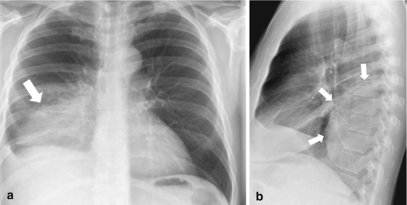
Cxr loculated right pleural effusion. A variety of fluids can be involved in left pleural effusion, including blood, pus from infections, lymph, and serous fluids. A ct study revealed this to be a loculated pleural effusion. Cxr = chest x ray; What are the different appearances of pleural effusion?
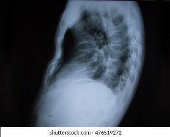
Loculated Pleural Effusion Images Stock Photos Vectors Shutterstock from image.shutterstock.com Loculated right pleural effusion with foci of atelectasis and consolidative changes concerning for pneumonia. Loculation most commonly occurs with exudative fluid, blood and pus. Surgical thoracostomy tube placement and radiologically guided catheter drainage are standard therapy for loculated pleural fluid collections. Pleural fluid glucose < 60 mg/dl; Line not corresponding to fissure A ct study revealed this to be a loculated pleural effusion. Enlarged mediastinal lymph nodes, possibly reactive. The fluid is locked in place despite gravity.
The first step in evaluating pleural effusions is determining whether it is transudative or exudative. Normally, a small amount of fluid is present in the pleura. What are the different appearances of pleural effusion? Complex septated, complex nonseptated, or homogeneously echogenic effusions are always exudates (fig. Sometimes in the setting of pleuritis, loculation of fluid may occur within the fissures or between the pleural layers (visceral and parietal). Most patients with pleural malignancy, either primary or secondary, develop pleural effusions. A variety of fluids can be involved in left pleural effusion, including blood, pus from infections, lymph, and serous fluids. Treatment may fail if the catheter is not placed optimally within the loculation or if the fluid is hemorrhagic or fibrinous. This type of effusion is empyema unless proven otherwise. Icu patients cannot sit up and the effusion layers posteriorly. Cxr = chest x ray; Differential diagnosis of pleural effusion; Most effusions start like this and can be easily missed.